Technological progress in visualisation techniques like 3D print support clinics in developing customised surgical solutions for patients. Due to the increasing development of technologies and, thus, more complex surgical interventions, better surgery preparation, digital planning and further qualification of doctors are needed. Especially for pre-operation planning 3D models can help create realistic and customised pathologies of the anatomic regions affected and support at therapy decision, or they help visualise the actual situation to doctors-to-be and give them the possibility to simulate the operation in advance.
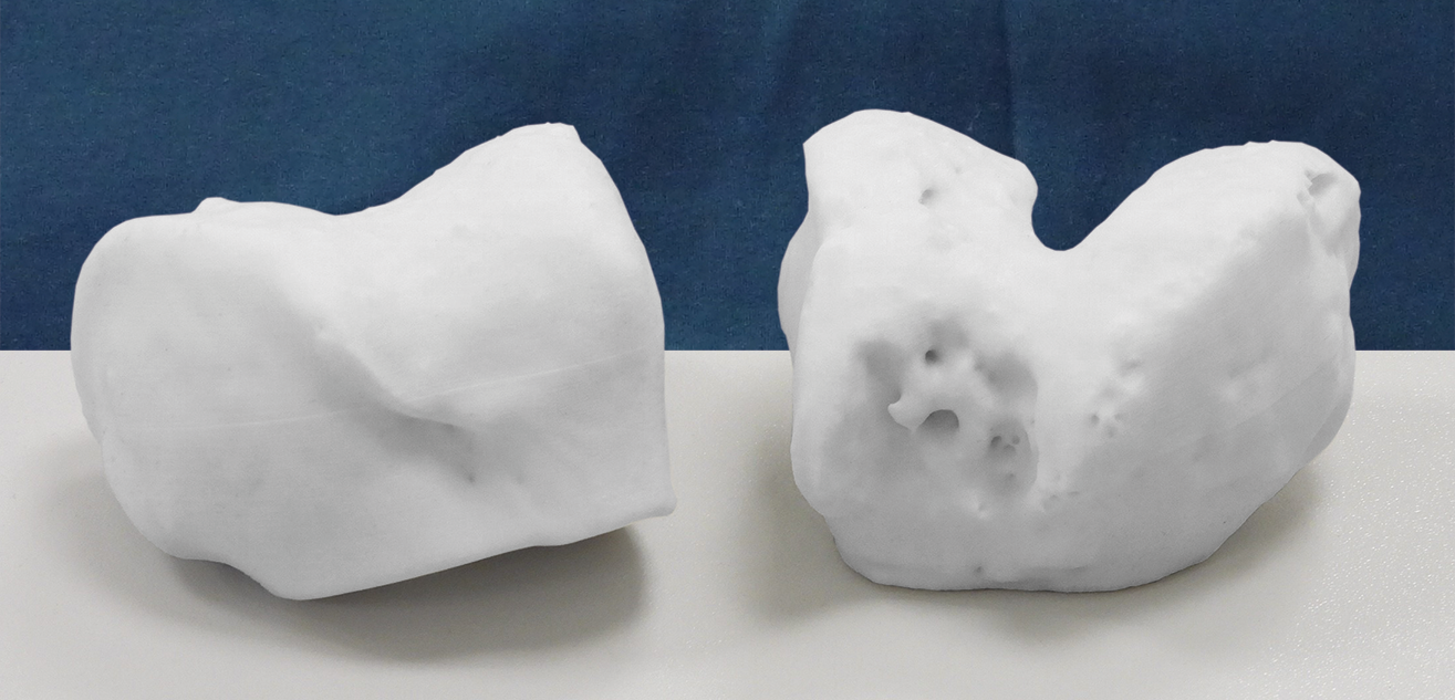
In this case, the patient has a healthy trochlea at her right leg and a valgus malposition in comination with antetorsion of the femur at her left leg. Additionally, the patient was treated with two failed trochleoplasties which caused a necrosis in this area. Therefore, the attending doctor had the idea to take the healthy side as a model for the pathological side in order to get an adequate possibility to prepare the trochleoplasty. He ordered a 3D print for both trochleae to get a comparison of both and to simulate the operation at the affected trochlea.
For this purpose, he used the mediCAD® 3D Knee Software to segment the area of a CT which he wanted to 3D print and exported the model as .stl file. Directly from the mediCAD® user interface he opened the mediCAD® service portal for the order process. At this portal he received a price quote for the 3D print. Then everything went really quickly: The doctor entered his delivery address and the print was at his desk only 4 work days later.
3D print of the distal femur to prepare a trochleoplasty
Young female patients suffer quite often from pain in their patella caused by a valgus in combination with an augmented antetorsion of the femur. This was also the case of a young patient who went into this clinic. We ask the attending doctor about his experience.
Case Study

mediCAD®: Which treatment options do you see for this young female patient?
Doctor: The patient is very young and we have to very carefully reflect and plan what we can “do to her“ surgically in this young age.
mediCAD®: Previous conservative therapies have failed?
Doctor: Yes, and there were two failed trochleoplasties because osseous deformities like a pathological TT-TG, a valgus, especially at the left leg, haven‘t been considered. Instead of straightening the leg axis, the therapy was reduced to the trochlea hole.
mediCAD®: What can be done better in the case of such a young patient?
Doctor: Generally, for this kind of pain at the patella the range of treatments includes MPFL-reconstruction, tuberosity transfer, trochleoplasty and osteotomies to correct the leg axis and the torsion. Often, we choose combined interventions. In this case, we decided on a medial opening wedge HTO at the left side to get symmetry of both legs, thus changing also the patella tracking by cutting above the starting point of the patella sinew, the tuberosity tibiae. We start always with interventions of axis correction before pursuing changes of the trochlea or the ligaments.
mediCAD®: That sounds fascinating. Was this enough to release the patient free of pain from the hospital?
Doctor: We had corrected only one leg and wanted to see first how she was doing with it - even though we knew already that the trochleae was very dysplastic, or was affected by two trochleoplasties which were not working satisfactorily. The decision for the intervention had been taken at a follow-up examination. Most importantly, we now had to do everything right and plan the intervention in detail. Therefore, I wanted to use two 3D prints because I believe that trochleoplasty can be planned very well with this tool.
mediCAD®: Perfect, the future will certainly offer new opportunities for the planning of a trochleoplasty.
Doctor: I agree! In my eyes, 3D planning in combination with the possibility of 3D print will be the future of sugery planning.
mediCAD®: Thank you very much for the interview and your time!

